Hysteroscopy Before IVF: When Is It Needed?
Learn when hysteroscopy may be recommended before IVF, what uterine problems it can detect or treat, and how it may help prepare the uterus for embryo transfer.
Hysteroscopy before IVF sits in an awkward middle ground. It is genuinely valuable for some women and unnecessary for others, and the honest position is that it should not be routine for everyone. The question worth answering is not whether hysteroscopy works, but whether you specifically need one. Here is how that decision should be made.
What Hysteroscopy Actually Is
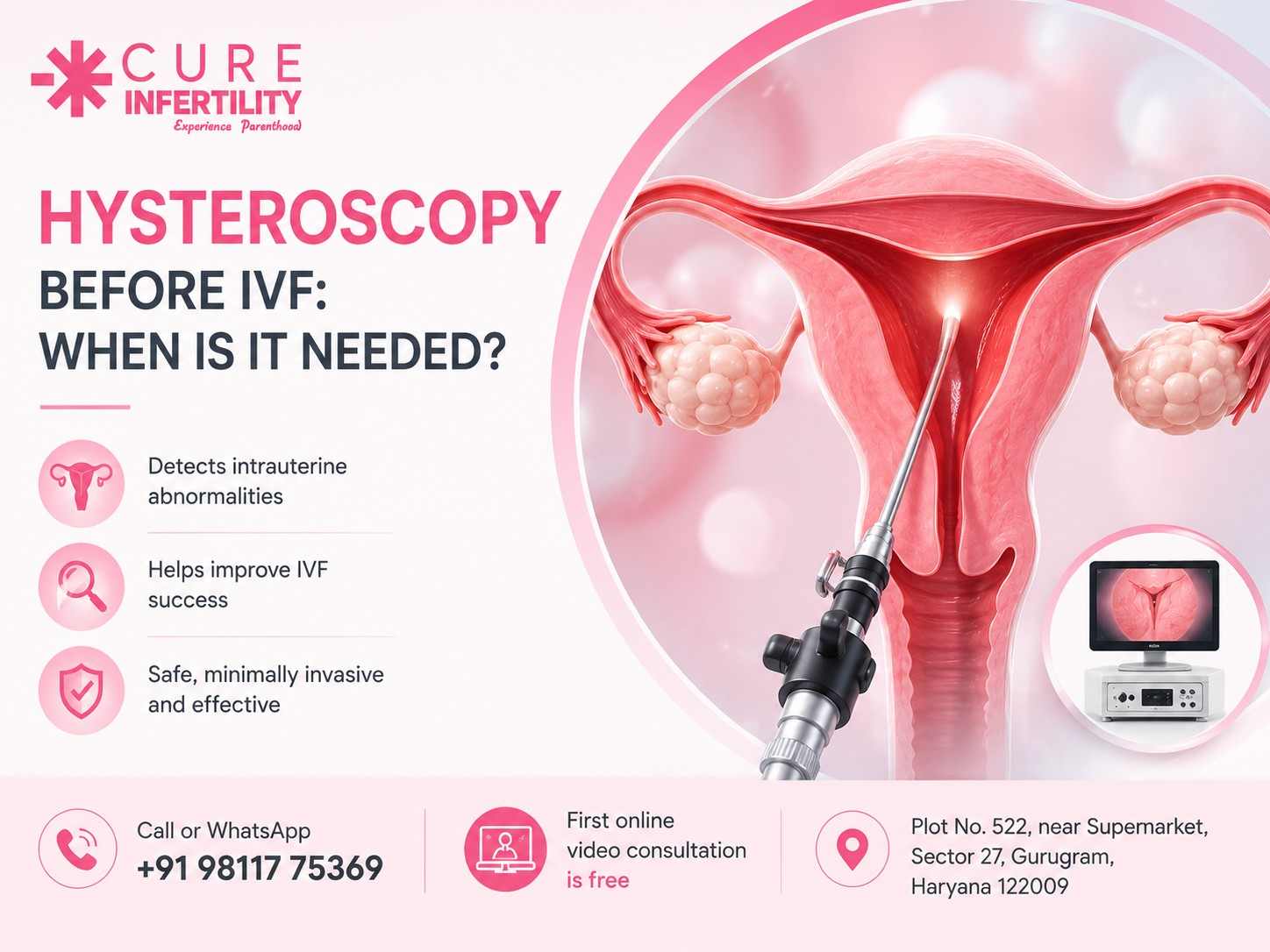
A hysteroscope is a thin telescope passed through the cervix into the uterine cavity, giving a direct view of the inside of the uterus on a screen. No incision is involved, since the natural passage of the cervix is used.
There are two versions, and the distinction matters.
Diagnostic hysteroscopy is a look only. It is often done in an outpatient setting, sometimes without anaesthesia or with local anaesthesia, and takes a matter of minutes.
Operative hysteroscopy uses a slightly wider scope with channels for instruments, so that anything found can be treated in the same sitting. This is usually done under general or regional anaesthesia as a day procedure.
The practical advantage of the second is significant. Where a polyp or adhesion is found, it is dealt with immediately rather than requiring you to return for a second procedure weeks later, which in an IVF timeline can mean an entire lost cycle.
Why the Uterine Cavity Matters So Much in IVF
An IVF cycle can produce an excellent embryo and still fail if the cavity that embryo is placed into is compromised. Implantation depends on the embryo making contact with a healthy, receptive endometrial surface.
A polyp sitting in the wrong position, a fibroid pushing into the cavity, or a band of scar tissue can prevent that contact entirely, or create an environment where implantation is far less likely. None of these produce symptoms in most women. You can have a completely normal cycle history and a compromised cavity.
That is the core argument for looking before transfer rather than after failure.
What Hysteroscopy Can Find
Endometrial polyps. Common, frequently missed on routine ultrasound, and straightforward to remove. Even small polyps in certain positions can interfere with implantation.
Submucosal fibroids. Fibroids that push into the uterine cavity have a clearly demonstrated negative effect on implantation, unlike fibroids sitting within the wall or on the outer surface, which often need no intervention at all. Location matters far more than size here.
Intrauterine adhesions (Asherman's syndrome). Bands of scar tissue, usually following a previous D&C, uterine surgery or infection. These can range from mild filmy bands to a cavity that is substantially obliterated. Symptoms may include lighter or absent periods, though many women have none.
Uterine septum. A congenital band of tissue dividing the cavity, associated with both implantation failure and recurrent miscarriage. Correction is done hysteroscopically.
Chronic endometritis. Low-grade inflammation of the lining that causes no symptoms and does not show on ultrasound. Hysteroscopy can show suggestive appearances, and a biopsy taken at the same time confirms it. It is treatable with antibiotics, which makes missing it particularly costly.
Cervical canal problems, including stenosis or an unusual angle, which can make embryo transfer technically difficult. Knowing this in advance allows the transfer to be planned rather than improvised on the day.
Why Ultrasound Is Not Enough
This is the crux of the argument, and it is worth being specific.
A standard transvaginal ultrasound is good at some things and poor at others. It reliably identifies larger fibroids and endometriomas. It is considerably less reliable for small polyps, thin adhesions, mild septa and anything involving the surface appearance of the endometrium.
Saline infusion sonography improves on plain ultrasound by distending the cavity with fluid, and it is a reasonable screening step. But it still shows a shadow rather than a direct view, and it cannot treat what it finds.
Hysteroscopy is the reference standard because it looks directly at the surface, and because it can act on what it sees in the same sitting.
Who Should Have One Before IVF
There is reasonable consensus that hysteroscopy is worthwhile in these situations:
- After one or more failed IVF cycles, particularly where good-quality embryos were transferred without success. This is the strongest indication, and it forms part of the wider workup described in our article on tests to consider after repeated IVF failure.
- Where ultrasound or saline sonography suggests an abnormality, to confirm and correct it.
- A history of recurrent miscarriage, where a septum or adhesions may be contributing.
- Previous uterine surgery, including D&C, myomectomy or caesarean section, all of which carry a risk of adhesion formation.
- Abnormal bleeding patterns, including unusually light periods, which can indicate adhesions.
- A known or suspected fibroid where its relationship to the cavity is unclear.
- A previous difficult embryo transfer, where cervical anatomy caused problems.
- Suspected chronic endometritis, particularly after unexplained implantation failure.
Who Probably Does Not Need One
Equally important, and less often said.
For a woman entering her first IVF cycle with a normal ultrasound, no relevant surgical history, regular periods and no suggestive symptoms, routine hysteroscopy is not clearly justified. Large studies looking at routine hysteroscopy before a first IVF cycle in women with a normal cavity on imaging have not shown a convincing improvement in live birth rates.
That is a meaningful finding, because hysteroscopy is not free, not entirely without risk, and adds time to a treatment timeline where time matters. Doing it as a default for every patient is difficult to defend clinically.
If it is being recommended to you, the useful question is which of the indications above applies in your case. A clear answer is a good sign. "We do it for everyone" is worth probing.
What the Procedure Involves
Timing. Usually scheduled in the first half of the cycle, after menstruation ends and before ovulation, when the lining is thin and the view is clearest.
The procedure itself. Diagnostic hysteroscopy typically takes five to ten minutes; operative procedures somewhat longer depending on what is being treated. The cervix is gently dilated where necessary, the scope is passed, and the cavity is distended with fluid to allow a clear view.
Anaesthesia. Diagnostic procedures may be done without anaesthesia or under local anaesthesia. Operative procedures are usually done under general or regional anaesthesia as a day case.
Afterwards. Mild cramping and light spotting for a few days is normal. Most women return to normal activity within a day or two, though recovery after a more extensive operative procedure takes a little longer.
Risks, which are low but real: infection, bleeding, and uncommonly perforation of the uterus. Fluid overload is a rare complication of longer operative procedures. These are worth understanding rather than fearing, and they are part of why the procedure should have a clear indication.
When Hysteroscopy Is Combined With Laparoscopy
Hysteroscopy examines the inside of the uterus. It cannot see the outside of the uterus, the ovaries, the fallopian tubes or the pelvic peritoneum.
Where the suspected problem lies outside the cavity, endometriosis, adhesions around the tubes and ovaries, or hydrosalpinx, laparoscopy is needed alongside it. The two are frequently combined in a single anaesthetic, which is efficient for the patient and gives a complete picture of both the cavity and the pelvis. Our article on endometriosis and fertility covers why this matters where endometriosis is suspected.
Hydrosalpinx deserves a particular mention. A fluid-filled blocked tube leaks fluid into the uterine cavity that is directly toxic to embryos, and treating it before transfer has good evidence behind it. This is picked up laparoscopically, not hysteroscopically.
How Long to Wait Before Starting IVF
After a straightforward diagnostic hysteroscopy with no intervention, an IVF cycle can usually begin in the next cycle.
After operative treatment, the interval depends on what was done. Polyp removal generally allows a short wait. More extensive work, such as adhesion division or septum correction, requires longer for the lining to heal properly, and sometimes a follow-up look to confirm the cavity has healed well and that adhesions have not reformed.
Your doctor should give you a specific interval based on the findings, not a generic answer.
The Honest Summary
Hysteroscopy is one of the most useful diagnostic tools in fertility medicine, and it has a real advantage in being able to treat as well as diagnose. Where an indication exists, particularly after failed cycles or with a relevant history, it is well worth doing.
Where no indication exists, in a first cycle with normal imaging and no relevant history, the case for it is weak, and the honest advice is that it can be skipped.
The route through IVF differs for everyone, and what matters is that each step in your plan has a reason attached to it. You can read more on the hysteroscopy and laparoscopy and IVF treatment pages, and see the full range of treatment options here.
You can book a free consultation with Dr. Rashmi Agrawal at the Sector 27, Gurugram clinic to review your imaging and history, read more about her background and approach, or check the FAQ page.
FAQs
Is hysteroscopy necessary before every IVF cycle?
No. It is clearly indicated after failed cycles, with abnormal imaging, a relevant surgical history or recurrent miscarriage. For a first cycle with a normal ultrasound and no relevant history, routine hysteroscopy has not been shown to improve live birth rates.
Is hysteroscopy painful?
Diagnostic hysteroscopy causes cramping similar to period pain in most women, and is often done without general anaesthesia. Operative hysteroscopy is done under anaesthesia, so it is not painful at the time, with mild cramping afterwards.
Can an ultrasound detect everything a hysteroscopy can?
No. Ultrasound reliably identifies larger structural problems but frequently misses small polyps, thin adhesions and mild septa, and it cannot detect chronic endometritis or treat anything it finds.
How soon after hysteroscopy can I start IVF?
After a diagnostic procedure with no intervention, usually the next cycle. After operative treatment, the interval depends on what was done, ranging from a short wait after polyp removal to longer after adhesion or septum correction.
Does removing a polyp improve IVF success?
Removing polyps that sit within the cavity is generally considered worthwhile, since they can interfere with implantation, and removal is straightforward with a short recovery.
Do all fibroids need to be removed before IVF?
No. Fibroids that push into the uterine cavity have a clear negative effect and are usually treated. Those within the wall or on the outer surface often need no intervention. Location matters more than size.
Can hysteroscopy detect endometriosis?
No. Hysteroscopy examines only the inside of the uterus. Endometriosis affects the pelvis outside the uterine cavity and requires laparoscopy to diagnose, which is why the two procedures are often combined.
What is chronic endometritis and why does it matter?
It is a low-grade inflammation of the uterine lining that causes no symptoms and does not appear on scans. It requires a biopsy to confirm and is treatable with antibiotics, making it one of the more valuable findings after unexplained implantation failure.
Medically reviewed
Reviewed by Dr. Rashmi Agrawal — MBBS (Gold Medalist), MS OBGYN, FNB Reproductive Medicine. This article is general information, not a substitute for a consultation about your own reports.
Book Consultation